UV Lights and Fluorescent Minerals - a fun side hobby to metal detecting !

The Battery and Bulb store in Little Rock had never seen this battery before and it was not listed in their computer! However, KT did find another US dealer on eBay with this same number battery, but under a different name….likely made by the same Chinese factory. KT ordered a pair from them and they are on their way from California. His Majesty will let e1 know when these come in and if they work and charge! BTW, these batteries cost around $7 US vs. the defective ones were $10 ! So 30% less.
So here is the update on the second set of new batteries for the SW flashlight. First they are almost the same, but instead of being button top, they are flat with the button on the + end flush….so…a little careful prying got each button up about 1 mm. They were put on the charger and charged just fine in a few hours. Then they were put in the flashlight and only worked intermittently. KT noticed that they were slightly rattley in the unit, so He searched through his Royal Collection of foreign coins, and found a small copper coin, inserted in between the two batteries and YES, the light now works! And the rattle was eliminated so all is well now. KT has now recharged the original batteries and has the spare set he desired in the first place! :kingdances:
 
Two fluorescent specimens arrived in the Royal Mailbox today all the way from Europe! Collected in the USA, sold or traded to a couple of European dealers and now back in the USA! :laughing:

The first is a massive, fibrous form of fluoborite, originally labeled from the Hopin’ #1 mine, but now labeled from the New Method mine, Amboy, San Bernardino Co., California. A small miniature. The first picture is in natural light, second picture is in LW 365nm, and third picture is in SW 254nm….an interesting massive specimen.

The second specimen consists of a crust of yellow Strelkinite crystals scattered on matrix from the Yellow Cat Mine, Thompson District, Grand Co., Utah. The crystals are micros on a T/N matrix. The first picture is in natural light, see the scale. The second picture is in LW 365nm and reveals that all the yellow crust is not strelkinite as only a portion of the yellow crust fluoresces!

All these photos were taken with KT's Royal Google Pixel 7 phone camera, focus is pretty decent, especially in both LW and SW UV. And using a newly purchased Cm/mm ruler!

Enjoy the pictures!
 

Attachments

  • Fluorborite, Hopin' #1 mine, New Method Mine, Amboy, San Bernardino Co., CA, natural light.jpg
    Fluorborite, Hopin' #1 mine, New Method Mine, Amboy, San Bernardino Co., CA, natural light.jpg
    282.1 KB · Views: 42
  • Fluorborite, Hopin' #1 mine, New Method Mine, Amboy, San Bernardino Co., CA, LW 365nm.jpg
    Fluorborite, Hopin' #1 mine, New Method Mine, Amboy, San Bernardino Co., CA, LW 365nm.jpg
    217 KB · Views: 40
  • Fluorborite, Hopin' #1 mine, New Method Mine, Amboy, San Bernardino Co., CA, SW 254nm.jpg
    Fluorborite, Hopin' #1 mine, New Method Mine, Amboy, San Bernardino Co., CA, SW 254nm.jpg
    154.6 KB · Views: 42
  • Strelkinite , Yellow Cat Mine, Thompson District, Grand Co., Utah, natural light.jpg
    Strelkinite , Yellow Cat Mine, Thompson District, Grand Co., Utah, natural light.jpg
    198.2 KB · Views: 42
  • Strelkinite , Yellow Cat Mine, Thompson District, Grand Co., Utah, LW 365nm.jpg
    Strelkinite , Yellow Cat Mine, Thompson District, Grand Co., Utah, LW 365nm.jpg
    163.5 KB · Views: 43
The first specimen is a fossil Stromatolite from the Anti-Atlas Mountains of eastern Morocco. The formation it was collected from has been age dated at 600 million years old, placing it in the Neoproterozoic Era of the Precambrian. Stromatolites consist of filamentous algae, and as so make up some of the worlds oldest life forms. Some have been dated to as early as 3 billion years before present! The rock consists of iron oxide-stained calcite cemented sandstone. The calcite cement is what fluoresces. The first image is in natural light and the second is in LW 365nm.

The second specimen consists of a mixture of Aragonite and Chondrodite in a Calcite matrix from Long Lake Zinc Mine, Parham, Olden Twsp., Frontenac County, Ontario, Canada. The first picture is in natural light and gives no hint of the variety of mineralization, just looking like a calcite marble. But the second picture in SW 254nm gives a kaleidoscope of colors...Aragonite responds as blue white (termed lightning bolt by some collectors), Chondrodite responds with yellow, and the matrix calcite is a nice red. Pictures are in excellent focus! This last image was taken using 3 SW lamps, one from bottom of image(50+ year old UV Products), one from the left side(11 watt tube) and the other from the top of the image at 45 degrees (the SW flashlight).

Enjoy these treats from nature’s fluorescent candy bowl!
 

Attachments

  • Fossil Stromatolite, Anti-Atlas Mountains, eastern Morocco, natural light.jpg
    Fossil Stromatolite, Anti-Atlas Mountains, eastern Morocco, natural light.jpg
    227.6 KB · Views: 41
  • Fossil Stromatolite, Anti-Atlas Mountains, eastern Morocco, LW 365nm.jpg
    Fossil Stromatolite, Anti-Atlas Mountains, eastern Morocco, LW 365nm.jpg
    209.7 KB · Views: 41
  • Aragonite & Chondrodite in Calcite, Long Lake Zinc Mine, Parham, Olden Twsp., Frontenac Co., O...jpg
    Aragonite & Chondrodite in Calcite, Long Lake Zinc Mine, Parham, Olden Twsp., Frontenac Co., O...jpg
    178.2 KB · Views: 46
  • Aragonite & Chondrodite in Calcite, Long Lake Zinc Mine, Parham, Olden Twsp., Frontenac Co., O...jpg
    Aragonite & Chondrodite in Calcite, Long Lake Zinc Mine, Parham, Olden Twsp., Frontenac Co., O...jpg
    112.1 KB · Views: 48
This is a form of chalcedony that KT has heard referred to as a Chalcedony Rose. This example is from the North Slope of Saddle Mountain, west of Phoenix, Maricopa County, Arizona. This specimen KT is giving to a local friend who also collects minerals, Stephanie Blandin, because she likes fluorescent minerals also. This was in a bag of goodies given to His Majesty by Jim Stoops! Thanks, Jim!

The fluorescence is due to the incorporation of a trace of Uranium during formation of this variety of quartz.

The first picture is in natural light and the second in LW 365nm. Taken with my Google Pixel 7 phone camera!
 

Attachments

  • Chalcedony, North Slope, Saddle Mtn., west of Phoenix, Maricopa Co., AZ, natural light.jpg
    Chalcedony, North Slope, Saddle Mtn., west of Phoenix, Maricopa Co., AZ, natural light.jpg
    222.2 KB · Views: 41
  • Chalcedony, North Slope, Saddle Mtn., west of Phoenix, Maricopa Co., AZ, LW 365nm.jpg
    Chalcedony, North Slope, Saddle Mtn., west of Phoenix, Maricopa Co., AZ, LW 365nm.jpg
    195 KB · Views: 43
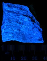
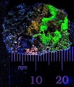
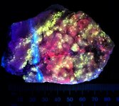
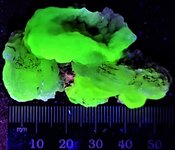
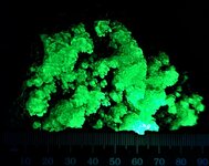
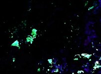
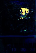
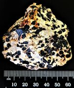
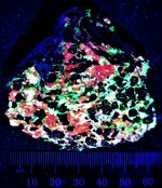
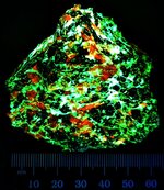
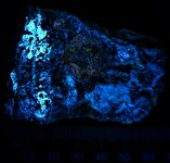
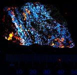
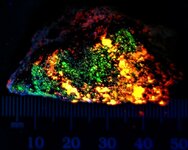
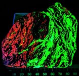
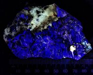
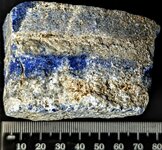
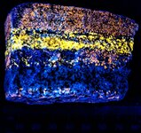
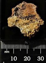
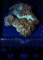
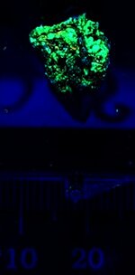
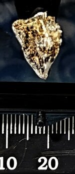
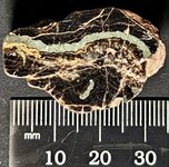
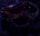
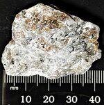
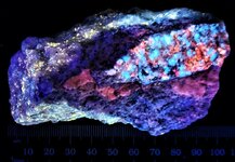
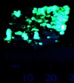
FYI – you might have noticed that the Royal pictures, especially those of minerals fluorescing, have greatly improved over the past several posts, and if you have not, then go back and look at the photos in the last 5 posts in this thread! :lol: :lol:

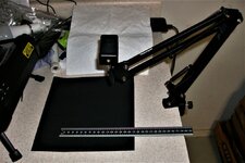
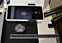
Anyway, KT wanted to document for you what has changed. Now instead of the Royal digital camera being put on a tripod to take the photos, KT is taking them with the camera on the Royal Google Pixel 7 phone. KT first tried taking a few by holding the camera and they were out of focus. Then He noticed that the GP7 camera is so sensitive to movement that it was constantly refocusing on the object, so…..His Majesty purchased from eBay a stable swing arm clamp unit with a mount for The Royal Cell phone. This was mounted on a cabinet, and KT covered the counter top, which was cream colored, with a black sheet of art paper. Mounted the cell phone, stuck a specimen in the field of view, snapped a picture, and discovered that the paper was actually dark purple to the camera. So the paper got a coat of flat black rattle can spray paint! After drying, now it looks black to the GP7 camera.

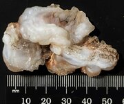
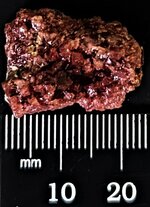
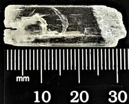
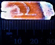
Then KT realized, that He had been taking pictures using US coins for scale. Good for some, but not for all His Majesty's email people, especially in Europe! And the coin made for odd shaped pictures. So, KT talked with the Queen and She showed Me some scales she had, but they were all in inches. KT searched online and found the company and they also sell scales with metric on 1 side and inches on the other. These are machinist scales and His Majesty picked the 1 foot/30 cm scale in black with white markings as it is more non-reflective than brushed steel with black markings. The scale is actually marked in cm with mm divisions on the side KT uses and below, out of the lower portion of the image, it is marked with 0.5 mm markings. The first picture shows the general set up of the paper, scale and phone clamp.

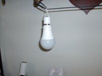
The second picture shows the phone held by the swivel arm clamp (you can read the clamp makers name on it), and a specimen on the paper, phone turned on, set for camera, and ready to take a picture. The lighting in the dungeon is overhead fluorescent fixtures, old ones with 4 bulbs. If KT does not like the lighting, He leaves the overheads on and turns on the 60 watt white light led bulb (picture 3) that is suspended from His mounted Royal moose antlers. His Majesty can easily slide it back and forth to get whatever directional shadowing effect He wants. The Queen had bought a pack of 4 of these rechargeable bulbs. They are for camping, and power outages, or whenever you just want to hang a light to use for a short time. They contain a Li ion battery in the base and you just screw them into any normal lamp receptacle and turn the light on to recharge them. Pretty neat set up. KT bought a pack of two of them just for his photo use. Good for 4 hours of continuous use, according to the manufacturer. The little black button on the top of the fixture is the switch.

The nice thing about this set up, aside from the camera focus not being affected by UV light, is that HIs Majesty does not have to touch the phone to take a picture. If He hovers a Royal finger about 1 mm above the white dot (the camera button), then it snaps the photo! This eliminates the chance for vibration.

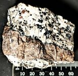
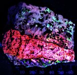
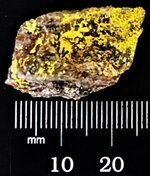
KT knows many people have cell phones and use their camera on them all the time, but the pictures they often take are portraits to scenic in nature, not pics of rocks and their fluorescence! But this change of equipment and technique appears to have resolved His Majesty's poor focus problems, particularly with UV lighting! BTW, these 3 pictures were taken with The Olde Royal digital camera and work just fine for what He is trying to show! So it will not go in the trash can until it dies.

Other than the cost of the cell phone, the Royal equipment costs for this set up was ~$20US for the camera swing arm clamp, and $10US for the scale. The Queen had the sheet of black construction paper, and KT had the can of spray flat black paint.
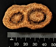
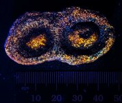
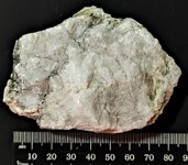
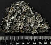
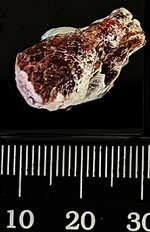
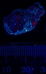
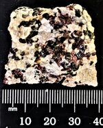
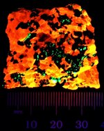
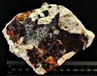
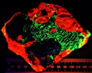
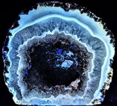
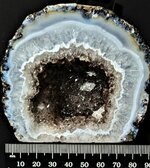
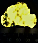
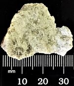
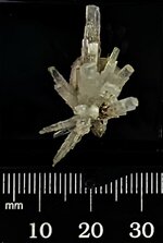
Enjoy the photos!
 

Attachments

  • Cell phone photo set up01.JPG
    Cell phone photo set up01.JPG
    109.9 KB · Views: 50
  • Cell phone photo set up02.JPG
    Cell phone photo set up02.JPG
    118.2 KB · Views: 41
  • Cell phone 2ndary light source.JPG
    Cell phone 2ndary light source.JPG
    49.3 KB · Views: 42
Several packages arrived today, including a new Royal Chinese USB led microscope, but for now here is one of the larger specimens.

A nice glassy coating of Opal (An)….while certainly not considered a mineral, opal is well known for its gem and fluorescent properties. This hyaline specimen consists of a nice bubbly coating of colorless opal on a black matrix of vesicular basalt from the Zemplen Mountains, Borsod-Abauj- Zemplen County, Hungary. This type of opal often fluoresces, having formed from very late water and often picks up traces of Uranium from those fluids.

The first picture is in natural light with the specimen on a black background. The second picture was taken using 2 SW 254nm UV lamps...KT's olde but Royal UV products 2 bulb model, and the newer 11-watt single tube unit, the former set at the base of the specimen and the latter on the left side of the image looking down at about 45 degrees….KT took another picture using both those lamps and the new Royal SW flashlight, but the picture was over exposed by the 3 light sources! This specimen does fluoresce weakly with LW 365nm, but no picture was taken.

There will be more pictures coming later this week! The Royal Family hopes everyone has a great and safe 4th of July Holiday tomorrow!

Enjoy the pictures!
 

Attachments

  • Opal (An) on vesicular basalt, Zemplen Mtns., Borsod-Abauj- Zemplen Co., Hungary, natural light .jpg
    Opal (An) on vesicular basalt, Zemplen Mtns., Borsod-Abauj- Zemplen Co., Hungary, natural light .jpg
    251.7 KB · Views: 42
  • Opal (An) on vesicular basalt, Zemplen Mtns., Borsod-Abauj- Zemplen Co., Hungary, SW 254nm.jpg
    Opal (An) on vesicular basalt, Zemplen Mtns., Borsod-Abauj- Zemplen Co., Hungary, SW 254nm.jpg
    115.9 KB · Views: 38
The first of several fluorescent micros I received today! The pictures were taken using my new Royal Chinese toy USB led microscope. The first one stripped an internal gear, likely plastic, so it could no longer be focused. It went in the trash can when this one arrived. Prince John assisted His Highness by uninstalling the previous Chinesium image software and then installed the newer Chinesium software that came with this unit. :laughing: :laughing:

After some tinkering around, KT got the it in focus and took a couple of test pictures for color rendition of colored letters on a printed piece of plastic. That worked so He took this specimen and got photographs of it.

The mineral of interest is Heinrichite, a hydrated barium uranium arsenate, which rapidly dehydrates, so likely it is already Meta-heinrichite. The specimen is from Krunkelbach Valley, Menzenschwand, Baden-Wurttemberg, Germany. Magnification is 10X. It is not very noticable in the natural light image, but once one sees the LW 365nm image...2nd image...one can see its distribution more readily on the natural light image. The third picture is also a LW 365nm image, where the light was held too close and over exposed the picture. KT did what He could to correct both the exposure and the color. The second image was taken with the UV LW lamp held some 6 inches from the specimen, whereas the 3rd picture it was ~ 1 inch from the specimen….way too close! Just wanted my Royal viewers to see what can happen with an over exposure in UV LW. HA HA

Enjoy the pictures! KT is really glad to have this new scope up and working!
 

Attachments

  • Heinrichite, Krunkelbach Valley, Menzenschwand, Baden-Wurttembere, Germany, 10X, LW 365nm 02.JPG
    Heinrichite, Krunkelbach Valley, Menzenschwand, Baden-Wurttembere, Germany, 10X, LW 365nm 02.JPG
    123.7 KB · Views: 37
  • Heinrichite, Krunkelbach Valley, Menzenschwand, Baden-Wurttembere, Germany, 10X, LW 365nm.JPG
    Heinrichite, Krunkelbach Valley, Menzenschwand, Baden-Wurttembere, Germany, 10X, LW 365nm.JPG
    60.8 KB · Views: 42
  • Heinrichite, Krunkelbach Valley, Menzenschwand, Baden-Wurttembere, Germany, 10X, natural light.JPG
    Heinrichite, Krunkelbach Valley, Menzenschwand, Baden-Wurttembere, Germany, 10X, natural light.JPG
    205.9 KB · Views: 39
Happy Independence Day!

First, KT has what was first a mystery specimen. It came labeled by the European dealer as Strelkinite, but after looking over the few pictures in Mindat.org, it was just not right. However, after a bit more looking, His Majesty came across pictures of Uranopilite that look essentially identical as this mineral and from the same Mining District! So He is calling it Uranopilite from the Yellow Cat Mining District, Grand Co., Utah. KT would have liked to pin the locality down a bit closer, however there are some 20 mines in that mining district, mostly for vanadium and/or uranium, and several have Uranopilite listed as present in Mindat.org. At least the district location by the dealer was reasonable.

The first picture is in natural light at 10X, and the mineral appears as spotty, blebby to platy olive-green grains scattered about on the matrix, which is mostly milky quartz with some late black MnOx. The specimen's overall size is a miniature with the fluorescent mineral as an encrustation. The second picture, surprisingly, appears in better focus! It was taken in LW 365nm and the lamp was held some 6 inches from the specimen. This mineral is also fluorescent in SW 254nm, but presents a weaker green response. In LW, it can be seen that the mineral is platy or at the least discoidal in habit.

The second mineral is Sal Ammoniac. KT purchased this specimen in the hope that it would be fluorescent, as listed in Henkel’s Glossary, but alas, it is not. However that did not stop Him from taking a photo of it as it is nicely crystallized. The specimen is from Sao Pedro da Cova, Gondomar, Porto, Portugal. Since Agricola’s time, this area has been mined for anthracite coal. Sal Ammoniac is a secondary mineral formed by the burning of coal waste piles from the mining operation. This mineral is highly water soluble so was recently formed before being collected. It occurs as both brown and colorless crystals and these are the typical forms as shown in several photos in Mindat.org. The picture was taken at 10X in natural light.

Enjoy the pictures and have a great Holiday! Hopefully, tomorrow’s mail will bring some additional specimens! I purchased several for my birthday, which was July 2nd, as presents to myself! :kingdances:
 

Attachments

  • Uranopilite, Yellow Cat Mining District, Grand Co., Utah, 10X, natural light.JPG
    Uranopilite, Yellow Cat Mining District, Grand Co., Utah, 10X, natural light.JPG
    137.6 KB · Views: 39
  • Uranopilite, Yellow Cat Mining District, Grand Co., Utah, 10X, LW 365nm.JPG
    Uranopilite, Yellow Cat Mining District, Grand Co., Utah, 10X, LW 365nm.JPG
    108.7 KB · Views: 35
  • Sal Ammoniac, NH4Cl, Sao Pedro de Cova, Gondomar, Porto, Portugal, 10X, natural light.JPG
    Sal Ammoniac, NH4Cl, Sao Pedro de Cova, Gondomar, Porto, Portugal, 10X, natural light.JPG
    180.5 KB · Views: 44
A bunch of arrivals in the Royal Mailbox today!

The first picture shows what was in the mailbox today, sans 1 piece of junk mail! HA HA Two envelops from Spain, box on left from Germany, and box on right from USA.

This posting documents two of the specimens. The first is Spinel from Sierra de Mijas, Mijas Malaga, Andalusia, Spain. The first picture is in natural light and the second is in LW. Small octahedral crystals.

The second specimen is of Hauyne from the Roca Negra Volcano, Santa Pau, Girona, Catalunya, Spain. The hauyne consists of pale blue grains intergrown with a light colored mineral on black scoria. The first picture is in natural light and the second is in LW 365nm, displaying the orange fluorescence typical of the Sodalite Group minerals.

Enjoy!
 

Attachments

  • Mail 07 05 2023 Top two are from Spain, lower left from Germany, and lower right from USA.JPG
    Mail 07 05 2023 Top two are from Spain, lower left from Germany, and lower right from USA.JPG
    118.1 KB · Views: 40
  • Spinel, Sierra de Mijas, Mijas, Malaga, Andalusia, Spain, natural light.jpg
    Spinel, Sierra de Mijas, Mijas, Malaga, Andalusia, Spain, natural light.jpg
    165.2 KB · Views: 39
  • Spinel, Sierra de Mijas, Mijas, Malaga, Andalusia, Spain, LW 365nm.jpg
    Spinel, Sierra de Mijas, Mijas, Malaga, Andalusia, Spain, LW 365nm.jpg
    140 KB · Views: 41
  • Hauyne, Rocxa Negra Volcano, Santa Pau, Girona, Catalunya, Spain, natural light.jpg
    Hauyne, Rocxa Negra Volcano, Santa Pau, Girona, Catalunya, Spain, natural light.jpg
    219.2 KB · Views: 40
  • Hauyne, Rocxa Negra Volcano, Santa Pau, Girona, Catalunya, Spain, LW 365nm.jpg
    Hauyne, Rocxa Negra Volcano, Santa Pau, Girona, Catalunya, Spain, LW 365nm.jpg
    136.5 KB · Views: 42
The first specimen featured in this posting is Calcite & Willemite with minor Zincite and Franklinite (NF) from Sterling Hill Mine, Franklin, Sussex Co., NJ. The first picture is in natural light, the second in LW 365nm and the third is in SW 254nm (using all 3 of my Royal SW lamps. Interesting color difference in the calcite from LW to SW UV, from red to orange. Willemite is well known from this locality to fluoresce strongly green in both wavelengths. And the minor areas of Zincite fluoresce a bright white, also in both LW and SW UV. Specimen is a large miniature.

The next specimen consists of Diopside and Phlogopite in marble from Gfohl, Krems-Land District, Lower Austria, Austria. The first picture is in natural light, and simply looks like a normal calcite marble. The second picture is in LW 365nm, the Diopside showing as white to cream colored grains and the 1 grain of phlogopite has a yellowish cast. This specimen is also a large miniature.

The last specimen is Sphalerite with Dolomite and minor zincite, Raubi Mines, Cave de Predil, Udine Province, Fruilivenizia, Glulia, Italy. The first image is in natural light. The second is in LW 365nm, the Sphalerite fluorescing a nice orange and the Dolomite a pale blue. The third image is in SW 254nm, and shows the zincite fluorescing a nice strong white. This specimen is also a large miniature.

Enjoy the pictures! Tomorrow is KT's Mexican Train Dominoes game day at His church, so His Majesty decided to go ahead and post these images this evening since He had time.
 

Attachments

  • Calcite, Zincite, Willemite with Franklinite (NF), Sterling Hill Mine, Franklin, Sussex Co., N...jpg
    Calcite, Zincite, Willemite with Franklinite (NF), Sterling Hill Mine, Franklin, Sussex Co., N...jpg
    209.5 KB · Views: 45
  • Calcite, Zincite, Willemite with Franklinite (NF), Sterling Hill Mine, Franklin, Sussex Co., N...jpg
    Calcite, Zincite, Willemite with Franklinite (NF), Sterling Hill Mine, Franklin, Sussex Co., N...jpg
    176.8 KB · Views: 41
  • Calcite, Zincite, Willemite with Franklinite (NF), Sterling Hill Mine, Franklin, Sussex Co., N...jpg
    Calcite, Zincite, Willemite with Franklinite (NF), Sterling Hill Mine, Franklin, Sussex Co., N...jpg
    200.6 KB · Views: 42
  • Diopside and Phlogopite in marble, Gfohl, Krems-Land District, Lower Austria, Austria, natural...jpg
    Diopside and Phlogopite in marble, Gfohl, Krems-Land District, Lower Austria, Austria, natural...jpg
    193.8 KB · Views: 45
  • Diopside and Phlogopite in marble, Gfohl, Krems-Land District, Lower Austria, Austria, SW 254nm.jpg
    Diopside and Phlogopite in marble, Gfohl, Krems-Land District, Lower Austria, Austria, SW 254nm.jpg
    88 KB · Views: 41
  • Sphalerite & Dolomite with minor zincite, Raubi Mines, Cave de Predil, Udine Province, Fruiliv...jpg
    Sphalerite & Dolomite with minor zincite, Raubi Mines, Cave de Predil, Udine Province, Fruiliv...jpg
    247.3 KB · Views: 41
  • Sphalerite & Dolomite with minor zincite, Raubi Mines, Cave de Predil, Udine Province, Fruiliv...jpg
    Sphalerite & Dolomite with minor zincite, Raubi Mines, Cave de Predil, Udine Province, Fruiliv...jpg
    215.2 KB · Views: 38
  • Sphalerite & Dolomite with minor zincite, Raubi Mines, Cave de Predil, Udine Province, Fruiliv...jpg
    Sphalerite & Dolomite with minor zincite, Raubi Mines, Cave de Predil, Udine Province, Fruiliv...jpg
    121.6 KB · Views: 41
Today there was only one new sample in the mailbox, but it is rather spectacular, in KT's Humble Opinion!

The specimen was sold as Microcline & Calcite from Sterling Hill Mine, Franklin, Sussex Co., NJ. But as you will see, there is another fluorescent mineral present!

The first picture displays what KT is calling the obverse side, like coin collectors do, for this is the only side photographed when the mineral was put up for sale. This side is shown first in natural light and then in the 2nd picture in SW 254nm (3 lamps used) and shows nicely the blue white response of Microcline and red orange response of Calcite, contrasting nicely. The 3rd picture shows the reverse of the specimen, first in natural light and then in the 4th image in SW 254nm. A difference of day and night! More dominant orange yellow calcite, green Willemite and only a trace of bluish white Microcline on the reverse.

KT likes the contrast between the 2 sides of the piece. It is a small miniature, but showy in UV!
 

Attachments

  • Microcline & calcite, Sterling Hill Mine, Franklin, Sussex Co., NJ, natural light obverse.jpg
    Microcline & calcite, Sterling Hill Mine, Franklin, Sussex Co., NJ, natural light obverse.jpg
    323 KB · Views: 41
  • Microcline & calcite, Sterling Hill Mine, Franklin, Sussex Co., NJ, SW 254nm obverse.jpg
    Microcline & calcite, Sterling Hill Mine, Franklin, Sussex Co., NJ, SW 254nm obverse.jpg
    224.6 KB · Views: 39
  • Microcline & calcite, Sterling Hill Mine, Franklin, Sussex Co., NJ, natural light reverse.jpg
    Microcline & calcite, Sterling Hill Mine, Franklin, Sussex Co., NJ, natural light reverse.jpg
    325.5 KB · Views: 41
  • Microcline & calcite, Sterling Hill Mine, Franklin, Sussex Co., NJ, SW 254nm reverse.jpg
    Microcline & calcite, Sterling Hill Mine, Franklin, Sussex Co., NJ, SW 254nm reverse.jpg
    186.1 KB · Views: 40
Today there was only one new sample in the mailbox, but the texture is interesting.

The specimen is Calcite and Willemite from the Sterling Hill Mine, Franklin, Sussex Co., NJ.

The first mineral picture displays the specimen in natural light and one will note what has been called flow structure banding. The 2nd picture in SW 254nm (3 lamps used) and displays a strong flow texture in both the Calcite and Willemite. Average specimens of these minerals from this locality typically have a n equigranular texture that resulted from the regional metamorphism of the zinc orebody, which this piece may have originally displayed, but due to faulting this texture has developed. This happened at a high enough pressure that flowage of the minerals occurred adjacent to the actual fault zone. One side of this specimen actually has an area of slickensides exposed, but no fluorescent minerals were smeared across that area. Geologists who are into tectonics would love this specimen! The piece is a fist sized specimen so will be put in my collection with the other larger pieces from this location.

This specimen is from the Charlie Butts Collection per the old labels. I included a small bit of his bio from Wikipedia below.

Charles Butts (1863–1946) was an American Paleontologist. In 1899 he got his bachlor’s degree, followed by a master’s in 1900 from Alfred University and in 1927 the University of Alabama awarded him an honorary doctorate of science. In 1901 he joined the USGS as an assistant geologist, and was promoted to paleontologist in 1909. In 1912 He became a geologist. He worked with the USGS until his retirement in 1933. Starting in 1933 he was hired by the Virginia Geological Survey, for mapping of the Paleozoic formations of the Appalachian Valley Region. While being so employed, he conducted a lot of geological surveys in other states including Alabama, Kentucky, and Virginia. He died on October 4, 1946 at the age of 83 in Washington D.C. and is buried at Fort Lincoln Cemetery in Brentwood, Maryland. The picture below is from 1901, courtesy of Wikipedia.

1688756738563.png
 

Attachments

  • Calcite & Willemite, fault zone, Sterling Hill Mine, Franklin, Sussex Co., NJ, natural light.jpg
    Calcite & Willemite, fault zone, Sterling Hill Mine, Franklin, Sussex Co., NJ, natural light.jpg
    302.9 KB · Views: 41
  • Calcite & Willemite, fault zone, Sterling Hill Mine, Franklin, Sussex Co., NJ, SW 254nm.jpg
    Calcite & Willemite, fault zone, Sterling Hill Mine, Franklin, Sussex Co., NJ, SW 254nm.jpg
    205.8 KB · Views: 40
Thanks to Bill Prior, a collecting buddy and former co-worker at the AGS (he is now also retired) for giving His Majesty the first specimen KT has to show today!

The first sample is a section of a fluorite-chalcedony-pyrite vein from the V-Intrusive, Garland County, Arkansas. This piece has been sawn and ground smooth but not polished. The first picture is in natural light and the second in LW 365nm. The fluorite displays an interesting texture and the chalcedony shows a yellowish cream response. KT had collected this site years ago while on a field trip and Bill likely collected this specimen at the same time. The actual vein contains some trace of gold, but it is unknown as to which mineral contains it. Charles Milton made thin sections of this rock and noted the presence of traces of brookite and other minerals present, also secondary sulfur crystals in tiny vugs formed by the weathering of pyrite.

The second sample is a raw example of Lapis Lazuli, with bands of Sodalite and Scapolite from Badakhshan, Afghanistan. The first image of it is in natural light, and the second in LW 365nm. Fine grained bands of yellow fluorescing Scapolite and orange fluorescing Sodalite are obvious in the UV image.

The third sample is a thumbnail specimen covered with micro crystals of Mimetite from Sante Eulalia, Chihuahua State, Mexico. KT purchased this because He had never seen fluorescent Mimetite. Best in Mid-wave, but also decent in LW 365nm, the first image is in natural light, and the second in LW 365nm.

The final specimen today is of Villiaumunite, that poisonous water soluble iodine mineral from Mont St. Hilaire. KT has one other specimen from the same location. It is purple in natural light...the first picture, and red in LW 365nm ….in the second picture. Not apparent in the natural light, there is significant alteration to this mineral, which dampens the fluorescent red response to irregular patches of unaltered mineral.

Anyway, enjoy the pictures today!
 

Attachments

  • Fluorite, Chalcedony, minor pyrite (NF), Vein, V Intrusive, Garland Co., AR, natural light.jpg
    Fluorite, Chalcedony, minor pyrite (NF), Vein, V Intrusive, Garland Co., AR, natural light.jpg
    157.7 KB · Views: 44
  • Fluorite, Chalcedony, minor pyrite (NF), Vein, V Intrusive, Garland Co., AR, LW 365nm.jpg
    Fluorite, Chalcedony, minor pyrite (NF), Vein, V Intrusive, Garland Co., AR, LW 365nm.jpg
    136.1 KB · Views: 44
  • Lapis with Scapolite & Sodalite, Badakhshan, Afghanistan, natural light.jpg
    Lapis with Scapolite & Sodalite, Badakhshan, Afghanistan, natural light.jpg
    270.4 KB · Views: 47
  • Lapis with Scapolite & Sodalite, Badakhshan, Afghanistan, LW 365nm.jpg
    Lapis with Scapolite & Sodalite, Badakhshan, Afghanistan, LW 365nm.jpg
    218.4 KB · Views: 37
  • Mimetite, Sante Eulalia, Chihuahua State, Mexico, natural light.jpg
    Mimetite, Sante Eulalia, Chihuahua State, Mexico, natural light.jpg
    115.8 KB · Views: 37
  • Mimetite, Sante Eulalia, Chihuahua State, Mexico, LW 365nm.jpg
    Mimetite, Sante Eulalia, Chihuahua State, Mexico, LW 365nm.jpg
    131.6 KB · Views: 39
  • Villiaumite, Mont St. Hilaire, Quebec, Canada, natural light.jpg
    Villiaumite, Mont St. Hilaire, Quebec, Canada, natural light.jpg
    195.3 KB · Views: 37
  • Villiaumite, Mont St. Hilaire, Quebec, Canada, LW 365nm.jpg
    Villiaumite, Mont St. Hilaire, Quebec, Canada, LW 365nm.jpg
    144.5 KB · Views: 43
One box with 2 specimens arrived today in the Royal Mailbox via the US postal service!

One KT purchased and the other a gift from the seller. First the gift…..

A rather stock equigranular miniature specimen of Calcite and Willemite with non-fluorescent Franklinite from the Sterling Hill Mine, Ogdensburg, Sussex County, New Jersey. The first picture is in natural light, and the second taken using all three of the Royal SW 254nm lamps. To the eye it looks fine in SW with only one lamp, but by using 3 lamps, it looks even better! :yes::lol:

The second specimen is the one KT purchased. This small cabinet specimen weighs in at a bit over 1 pound (477 g), and consists of coarse grained calcite, a few large non-fluorescent reddish brown Zincite grains, and an irregular mass of dark brown Tephroite as seen in the first picture(natural light). The second picture, taken in SW 254nm, holds the surprise! From the Tephroite has exsolved Willemite, having a nice green fluorescence. In this image, nearly all the Willemite is confined to the mass of Tephroite. KT likes the texture of this type of specimen as it displays something out of the ordinary in the cooling history of this example. See one of KT's earlier posts in this thread for more information, or look up "exsolved Willemite in Tephroite" via Google Search for further explanation. Since this specimen is a large fist sized/small cabinet piece, it will fit nicely with the larger specimens that Ed O’Dell so kindly gave His Majesty awhile back!

Anyway, enjoy the photos!
 

Attachments

  • Calcite, Willemite, & Franklinite (NF), Sterling Hill Mine, Ogdensburg, Sussex County, NJ, nat...jpg
    Calcite, Willemite, & Franklinite (NF), Sterling Hill Mine, Ogdensburg, Sussex County, NJ, nat...jpg
    204.1 KB · Views: 45
  • Calcite, Willemite, & Franklinite (NF), Sterling Hill Mine, Ogdensburg, Sussex County, NJ, SW ...jpg
    Calcite, Willemite, & Franklinite (NF), Sterling Hill Mine, Ogdensburg, Sussex County, NJ, SW ...jpg
    175.6 KB · Views: 44
  • Calcite, Tephroite with exsolved Willemite, and Zincite (NF), Sterling Hill Mine, Ogdensburg, ...jpg
    Calcite, Tephroite with exsolved Willemite, and Zincite (NF), Sterling Hill Mine, Ogdensburg, ...jpg
    200.3 KB · Views: 42
  • Calcite, Tephroite with exsolved Willemite, and Zincite (NF), Sterling Hill Mine, Ogdensburg, ...jpg
    Calcite, Tephroite with exsolved Willemite, and Zincite (NF), Sterling Hill Mine, Ogdensburg, ...jpg
    150.1 KB · Views: 43
One package today with 4 specimens arrived in the Royal Mailbox!

The first pair of pictures are of an Elpidite crystal cluster from the Poudrette Quarry, Mont St. Hilaire, Quebec, Canada. The first picture is in natural light and the second in LW 365nm.

The second pair of pictures are of a cluster of Natrolite from the Poudrette Quarry, Mont St. Hilaire, Quebec, Canada. The first of this pair is in natural light and the second is in LW 365nm.

The third pair of pictures are of a small miniature of Scapolite, Var. Wernerite, from Grenville, Quebec, Canada. The first picture of this pair is in natural light, and the second is in LW 365nm. To get a decent picture in LW that was not washed out, KT had to use a very weak LW lamp and hold it some 2 feet away from the specimen, due to the very strong UV response of this mineral!


The final pair of pictures are of a quartz crystal lined agate geode from British Columbia, Canada. KT knows, it looks more like a Mexican Coconut geode, but I only repeat what the label states! The first picture is natural light and the second is in LW 365nm. Note the small late calcite, fluorescing pale bluish white, perched on the quartz crystal lining! The agate fluoresces nicely in various shades of blue and blue white.

All these specimens came to me from a seller in Europe. Enjoy the pictures!
 

Attachments

  • Quartz crystal agate geode, British Columbia, Canada, LW 365nm.jpg
    Quartz crystal agate geode, British Columbia, Canada, LW 365nm.jpg
    195.8 KB · Views: 37
  • Quartz crystal agate geode, British Columbia, Canada, natural light.jpg
    Quartz crystal agate geode, British Columbia, Canada, natural light.jpg
    203.6 KB · Views: 42
  • Scapolite, var. Wernerite, Grenville, Quebec, Canada, LW 365nm.jpg
    Scapolite, var. Wernerite, Grenville, Quebec, Canada, LW 365nm.jpg
    125.1 KB · Views: 40
  • Scapolite, var. Wernerite, Grenville, Quebec, Canada, natural light.jpg
    Scapolite, var. Wernerite, Grenville, Quebec, Canada, natural light.jpg
    162.6 KB · Views: 42
  • Natrolite crystal cluster, Poudrette Qy., Mont St. Hilaire, Quebec, Canada, natural light.jpg
    Natrolite crystal cluster, Poudrette Qy., Mont St. Hilaire, Quebec, Canada, natural light.jpg
    126.7 KB · Views: 36
  • Elpidite crystal cluster, Poudrette Qy., Mont St. Hilaire, Quebec, Canada, LW 365nm.jpg
    Elpidite crystal cluster, Poudrette Qy., Mont St. Hilaire, Quebec, Canada, LW 365nm.jpg
    98.5 KB · Views: 38
  • Elpidite crystal cluster, Poudrette Qy., Mont St. Hilaire, Quebec, Canada, natural light.jpg
    Elpidite crystal cluster, Poudrette Qy., Mont St. Hilaire, Quebec, Canada, natural light.jpg
    126.3 KB · Views: 38
Today’s specimen is small, but interesting.

It consists of a polished example of Schalenblende (sphalerite) in a nugget of Galena, cut by a vein of Marcasite. The specimen was collected and polished by Albert Vercammen. Remembering back to my Royal college days, marcasite, like galena takes a high polish, but when galena is polished it is black whereas marcasite is surprisingly white or very light gray. So in natural light what you see most prominently in the black matrix is an almost translucent looking light colored veinlet of marcasite. The yellowish tan mineral is the sphalerite. Note that neither the marcasite nor the galena fluoresce, so in the second picture the small area of yellowish tan fluorescence is the sphalerite. Not a great specimen, but certainly interesting and from a locality I did not have (I have several nicer ones from Germany). The second picture was taken utilizing all 3 of the Royal SW 254nm lamps, placed in different positions around the specimen.

Enjoy the pictures! You will have to open the pictures to see what I am talking about since the fluorescence is not strong!
 

Attachments

  • Schalenblende with Marcasite vein in Galena nodule, Schmafgraf Mine, Liege, Wallonia, Belgium,...jpg
    Schalenblende with Marcasite vein in Galena nodule, Schmafgraf Mine, Liege, Wallonia, Belgium,...jpg
    200.8 KB · Views: 42
  • Schalenblende with Marcasite vein in Galena nodule, Schmafgraf Mine, Liege, Wallonia, Belgium,...jpg
    Schalenblende with Marcasite vein in Galena nodule, Schmafgraf Mine, Liege, Wallonia, Belgium,...jpg
    121.9 KB · Views: 43
Some interesting fluorescent minerals arrived today in the Royal mail!

First featured is a specimen of Wollastonite in Calcite from the Nobel Pit, Sterling Hill Mine, Ogdensburg, Sussex Co., New Jersey. The first photo is in natural light, just another gray white rock! HA HA. But the 2nd photo shows its response to SW 254nm UV. The calcite being an orangish red and the crystals of Wollastonite contrasting a golden yellow! This was a free gift from one of KT's favorite eBay dealers and was a good example for the Royal collection!

The next specimen is an interesting piece. The first photo shows some coarse dark Augite (originally called Jeffersonite) with a bit of calcite and willemite scattered about from Franklin Mine, Franklin, Sussex County, New Jersey. It has a different texture when responding to SW 254nm UV, that many of the specimens from this locality. The Willemite (green) is in some areas coarser grained and in others tiny specks scattered about, the calcite (orange red) is seen as patches of color scattered through the specimen. Then there is a mineral, not listed by the dealer, but which KT thinks is hydrozincite from its bright white response as grains cutting diagonally across the specimen! With the augite not fluorescing, it makes a nice black background for the other minerals to show off their fluorescent colors!

The third specimen was listed as Diopside on Calcite from the Long Lake Zn Mine, Parham, Olden Township, Frontenac County, Ontario, Canada. The first picture in natural light shows almost nothing but a whitish crystalline marble texture. But the SW 254nm fluorescent response reveals a section of a vein of diopside (bluish white) and patches of calcite (red). Fortunately the vein was broken through the thin vein instead of across it. The matrix also shows the presence of fine-grained Chondrodite (yellow orange speckles) throughout the matrix on one end of the specimen.

The final specimen today is spectacular in its own right! A Sphalerite vein is crosscuts a matrix of Calcite, Willemite, and Franklinite. The first picture is in natural light and the second is in LW 365nm. In that 2nd photo, the vein stands out orange in the pale matrix and the Willemite shows its distinctive green coloration. Note that some of the Sphalerite can be seen penetrating the matrix as small orangish grains in the 2nd image. KT loves being able to see these relationships under a UV light!

Enjoy the pictures!
 

Attachments

  • Wollastonite in calcite, Nobel Pit, Sterling Hill Mine, Ogdensburg, Sussex Co., NJ, natural li...jpg
    Wollastonite in calcite, Nobel Pit, Sterling Hill Mine, Ogdensburg, Sussex Co., NJ, natural li...jpg
    259.4 KB · Views: 40
  • Wollastonite in calcite, Nobel Pit, Sterling Hill Mine, Ogdensburg, Sussex Co., NJ, SW 254nm.jpg
    Wollastonite in calcite, Nobel Pit, Sterling Hill Mine, Ogdensburg, Sussex Co., NJ, SW 254nm.jpg
    147.8 KB · Views: 42
  • Willemite, Calcite, & Hydrozincite in Augite, Franklin Mine, Franklin, Sussex Co., NJ, natural...jpg
    Willemite, Calcite, & Hydrozincite in Augite, Franklin Mine, Franklin, Sussex Co., NJ, natural...jpg
    331 KB · Views: 38
  • Willemite, Calcite, & Hydrozincite in Augite, Franklin Mine, Franklin, Sussex Co., NJ, SW 254nm.jpg
    Willemite, Calcite, & Hydrozincite in Augite, Franklin Mine, Franklin, Sussex Co., NJ, SW 254nm.jpg
    224.5 KB · Views: 40
  • Diopside on Calcite with Chondrodite, Long Lake Zn Mine, Parham, Frontenac Co., Ont., Canada, ...jpg
    Diopside on Calcite with Chondrodite, Long Lake Zn Mine, Parham, Frontenac Co., Ont., Canada, ...jpg
    186 KB · Views: 39
  • Diopside on Calcite with Chondrodite, Long Lake Zn Mine, Parham, Frontenac Co., Ont., Canada, ...jpg
    Diopside on Calcite with Chondrodite, Long Lake Zn Mine, Parham, Frontenac Co., Ont., Canada, ...jpg
    124.5 KB · Views: 40
  • Sphalerite vein in Calcite & Willemite, Sterling Hill Mine, Ogdensburg, Sussex Co., NJ, natura...jpg
    Sphalerite vein in Calcite & Willemite, Sterling Hill Mine, Ogdensburg, Sussex Co., NJ, natura...jpg
    272.5 KB · Views: 38
  • Sphalerite vein in Calcite & Willemite, Sterling Hill Mine, Ogdensburg, Sussex Co., NJ, LW 365nm.jpg
    Sphalerite vein in Calcite & Willemite, Sterling Hill Mine, Ogdensburg, Sussex Co., NJ, LW 365nm.jpg
    192 KB · Views: 41
One package arrived today from Germany in the Royal Mailbox!

Turns out the contained fluorescent specimen made a trip around the world!:rofl2::rofl2::wow:

The mineral of interest is Zippeite (or perhaps Sodium-Zippeite; most analyzed specimens turn out to be according to Mindat.com) from the Parco #23 Mine, Yellow Cat Mesa, Thompsons Mining District, Utah, It went from that location, making its way to a German mineral dealer and then to me! Kinda a Christmas in July experience! :xtree::santa2:

Picture 1 shows yellow crusts of the Zippeite coating the host rock and Picture 2 shows the display of color under SW 254nm. Since this mineral is also highly fluorescent green in LW 365nm, you might wonder why no picture….well, even KT's weakest LW lamp caused such a bright response that His Majesty could not get a picture that was as well focused and green as with the SW lamps. No need therefore since the color response is the same, just as listed in Fluomin.org.

Enjoy the pictures!
 

Attachments

  • Zippeite, Parco #23 Mine, near Yellow Cat Mesa, Thompsons Mining District, Grand Co., Utah, na...jpg
    Zippeite, Parco #23 Mine, near Yellow Cat Mesa, Thompsons Mining District, Grand Co., Utah, na...jpg
    148.7 KB · Views: 41
  • Zippeite, Parco #23 Mine, near Yellow Cat Mesa, Thompsons Mining District, Grand Co., Utah, SW...jpg
    Zippeite, Parco #23 Mine, near Yellow Cat Mesa, Thompsons Mining District, Grand Co., Utah, SW...jpg
    92.1 KB · Views: 36
One package arrived in the Royal mailbox today!

A nice thumbnail specimen of Spodumene, a variety that not too long ago was termed Triphane, but now is considered an outdated varietal name (Mindat.org via the International Names Committee).

It is from the Dara-e-Pech pegmatite field, Dara-e-Pech District, Afghanistan. It measures slightly over 3 cm. The first picture is in natural light and the second in LW 365nm. The fluorescence is contained within a central zone of the crystal.

Enjoy the 2 pictures!
 

Attachments

  • Spodumene, var. Triphane, Dara-e-Pech Pegm. Field, Dara-e-Pech District, Kunar, Afghanistan, n...jpg
    Spodumene, var. Triphane, Dara-e-Pech Pegm. Field, Dara-e-Pech District, Kunar, Afghanistan, n...jpg
    155.3 KB · Views: 38
  • Spodumene, var. Triphane, Dara-e-Pech Pegm. Field, Dara-e-Pech District, Kunar, Afghanistan, L...jpg
    Spodumene, var. Triphane, Dara-e-Pech Pegm. Field, Dara-e-Pech District, Kunar, Afghanistan, L...jpg
    135.5 KB · Views: 37
Back
Top Bottom