Well, GKL, KT would love that but likely it will not happen. However, sometime opportunities arise. There is an 80 year old man in Kentucky who was a mining engineer for some 50+ years. He worked at major mining operations all over the central USA. He has now decided to sell his collection....200 specimens from many closed locations. A friend stopped in and looked over the collection and told me there is not one bad specimen in the entire collection. So we have got a group of 9 friends to kick in enough cash to meet his selling cost. Out of the group, KT is the only one that will not be present, but he is in it to get some specimens from some sites long closed, up to 50 years! If any are fluorescent, that will be sweet, but if not, KT will pick the favorites out of the 20+specimens He will get and sell the rest, likely to double the money invested. KT's friend is planning a trip down to AR in September, so he will store and then transport the specimens to the Castle during his visit! I am thinking some of these specimens may be worth over 100 bucks each, but will only know after His Majesty gets to examine them in detail! Now that is a deal! Sometimes it really pays to have connections!Neat KT, just maybe you might eventually get close to having a specimen from most of the countries in the world

You are using an out of date browser. It may not display this or other websites correctly.
You should upgrade or use an alternative browser.
You should upgrade or use an alternative browser.
UV Lights and Fluorescent Minerals - a fun side hobby to metal detecting !
- Thread starter GKL
- Start date
Several times KT has posted SW 254nm pictures and noted that there were 3 SW lamps set up to take the image. He thought perhaps since His Majesty only has 2 Royal hands, you might wonder how that is done and KT still be able to take the image. The first 4 pictures show how it is done. 
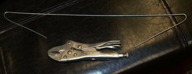
Picture 1 shows what was used to make a hanger for one of the lamps to be on the Royal tripod....
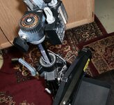
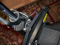
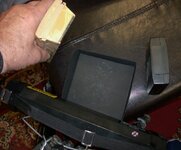
Picture 2 shows the position of the hanger on the tripod with the lamp hanging from it and where the camera mounts and the photo blackout box. Picture 3 shows the how the hanger is holding the lamp. And picture 4 shows how the other 2 SW lamps are set up when KT takes a picture. His Majesty thought this might interest someone.
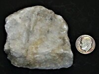
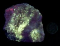
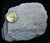
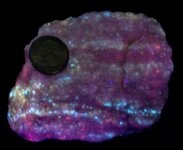
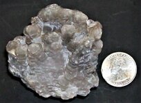
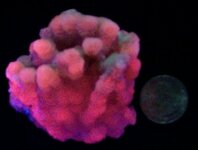
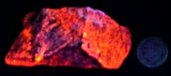
Picture 5 shows a natural light image of another calcite marble specimen from Long Lake Zn Mine, Ont., Canada. US dime in all pictures for scale. Note that in natural light no other mineralization can be spotted. Picture 6 shows the same specimen in the SW 254nm light from the 3 lights positioned as shown in picture 4. Note that Chondrodite fluoresces a nice yellow and the calcite matrix is pastel red.
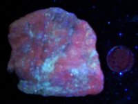
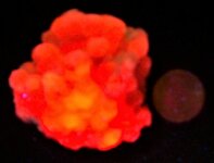
In Picture 6, taken in LW 365, chondrodite does not fluoresce, so the Diopside response is now visible, along with red responsive calcite. Just goes to show you always need to check with both LW and SW to get the more complete story! The specimen was sold with no mention of diopside on the specimen and no LW image provided.
Anyway, enjoy the pictures!
Picture 1 shows what was used to make a hanger for one of the lamps to be on the Royal tripod....
Picture 2 shows the position of the hanger on the tripod with the lamp hanging from it and where the camera mounts and the photo blackout box. Picture 3 shows the how the hanger is holding the lamp. And picture 4 shows how the other 2 SW lamps are set up when KT takes a picture. His Majesty thought this might interest someone.
Picture 5 shows a natural light image of another calcite marble specimen from Long Lake Zn Mine, Ont., Canada. US dime in all pictures for scale. Note that in natural light no other mineralization can be spotted. Picture 6 shows the same specimen in the SW 254nm light from the 3 lights positioned as shown in picture 4. Note that Chondrodite fluoresces a nice yellow and the calcite matrix is pastel red.
In Picture 6, taken in LW 365, chondrodite does not fluoresce, so the Diopside response is now visible, along with red responsive calcite. Just goes to show you always need to check with both LW and SW to get the more complete story! The specimen was sold with no mention of diopside on the specimen and no LW image provided.
Anyway, enjoy the pictures!
Attachments
-
 SW hanger01.JPG61.5 KB · Views: 60
 -
 SW hanger02 hanger on tripod.JPG171.4 KB · Views: 58
 -
 SW hanger03 holding SW 11 watt lamp.JPG132.8 KB · Views: 45
 -
 SW hanger04 how I take a SW picture with 3 SW lamps.JPG134.6 KB · Views: 50
 -
 Chondrodite, minor diopside, on calcite, Long Lake Zn mine, Parham, Olden Twsp., Frontenac Co....JPG129.4 KB · Views: 55
 -
 Chondrodite, minor diopside, on calcite, Long Lake Zn mine, Parham, Olden Twsp., Frontenac Co....JPG76.5 KB · Views: 55
 -
 Chondrodite, minor diopside, on calcite, Long Lake Zn mine, Parham, Olden Twsp., Frontenac Co....JPG94 KB · Views: 54
Should read in picture 7, taken in LW 365nm, chondrodite does not fluoresce.....Several times KT has posted SW 254nm pictures and noted that there were 3 SW lamps set up to take the image. He thought perhaps since His Majesty only has 2 Royal hands, you might wonder how that is done and KT still be able to take the image. The first 4 pictures show how it is done.
Picture 1 shows what was used to make a hanger for one of the lamps to be on the Royal tripod....
Picture 2 shows the position of the hanger on the tripod with the lamp hanging from it and where the camera mounts and the photo blackout box. Picture 3 shows the how the hanger is holding the lamp. And picture 4 shows how the other 2 SW lamps are set up when KT takes a picture. His Majesty thought this might interest someone.
Picture 5 shows a natural light image of another calcite marble specimen from Long Lake Zn Mine, Ont., Canada. US dime in all pictures for scale. Note that in natural light no other mineralization can be spotted. Picture 6 shows the same specimen in the SW 254nm light from the 3 lights positioned as shown in picture 4. Note that Chondrodite fluoresces a nice yellow and the calcite matrix is pastel red.
In Picture 6, taken in LW 365, chondrodite does not fluoresce, so the Diopside response is now visible, along with red responsive calcite. Just goes to show you always need to check with both LW and SW to get the more complete story! The specimen was sold with no mention of diopside on the specimen and no LW image provided.
Anyway, enjoy the pictures!


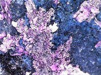
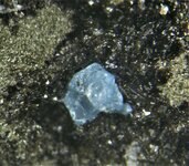
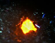
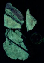
Here is a dangerous rare mineral! These photographs are of Villiaumite, NaF, with phlogopite from the Lovozero Massif, Murmansk Oblast, Kola Pennisula, Russia. This pair of images were taken with The Royal Chinese USB led microscope at 10X. The first photograph is in natural light with the Villiaumite showing its typical deep reddish purple color. The second photograph is in LW 365nm, showing its dark red fluorescence.
Villiaumite is associated with nepheline syenite (igneous rocks), particularly in gas cavities, so KT was somewhat surprised that it has not been reported from the Arkansas syenitic rocks. However, this is understandable when considering we do not have a dry climate. During the Paleocene/Eocene in Arkansas, our syenitic rocks were exposed to extensive lateritic weathering. Since this mineral is water soluble, that may explain why it has not been reported.
Health Risks of Villiaumite:
Poisonous! Fluoride ion in water-soluble form is what makes this mineral highly toxic. Its availability in macrospecimens, and the attractive colour, might lure small children into tasting it. May be fatal if swallowed or if dust is inhaled. Affects respiratory system, heart, skeleton, circulatory system, central nervous system and kidneys. Causes irritation to skin, eyes and respiratory tract. Irritation effects may be delayed. Rest assured that His Majesty washed His Royal hands after mounting this specimen. It is now encased in a plastic perky 1.25” cube! Health risk info from Mindat.org.
Attachments
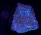
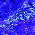
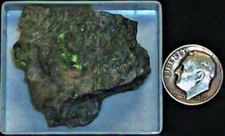
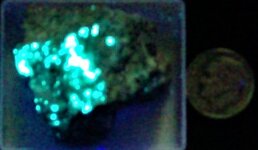
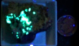
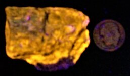
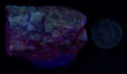
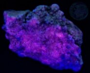
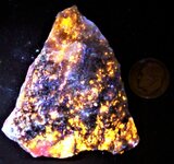
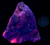
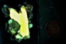
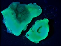
The first images today are of Hedyphane from the type locality of Langban Mine, Langban Ore District, Filipstad, Varmland Co., Sweden. The first image is in natural light, US dime for scale, second image is in SW 254nm, with all three lamps in the normal triangulated set up. The 3rd image is in LW 365nm, using the UVBeast at 3 feet and from the upper right side at a 45 degree angle. This is interesting because Mindat.org lists this mineral as not fluorescent in LW and being orange in SW. However, Fluomin.org lists several colors for the various wavelengths including even in mid-wave. I think Mindat.org for the fluorescent info is a bit out of date. That is exactly why I check several sources. Hedyphane is in the Apatite Group. As to what the blue-white responding mineral in the specimen in LW is, your guess is as good as KT's! 
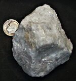
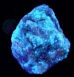
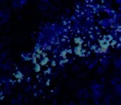
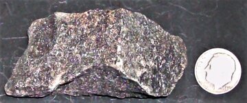
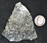
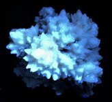
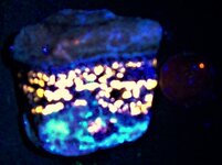
The second specimen has 3 pictures and is from the Long Lake Zinc Mine, Ontario, Canada. This piece has Chondrodite and Diopside set in Calcite. The first picture is in natural light, US quarter for scale. It appears to exhibit no banding in normal light, but in the second image in SW 254nm (3 SW lamps) it displays nice banding of the Chondrodite (yellow) and Diopside (blue-white) in the Calcite (deep red). The third picture is in LW 365nm, and since the Chondrodite has no response, the Diopside is more noticable in the deep red calcite matrix.
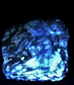
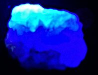
The last specimen today has 3 pictures and is also from Long Lake. The first picture is in natural light with a US dime for scale. This is what has been termed a “starry night” specimen. Little Chondrodite is present as revealed in the second picture, mostly Diopside in Calcite matrix, in SW 254nm (3 lamp set up). The last picture is in LW 365 and note how much stronger the red response of the Calcite is!
You might be getting tired of these Long Lake specimens but KT thinks they are most interesting! HA HA
Enjoy the pictures!
The second specimen has 3 pictures and is from the Long Lake Zinc Mine, Ontario, Canada. This piece has Chondrodite and Diopside set in Calcite. The first picture is in natural light, US quarter for scale. It appears to exhibit no banding in normal light, but in the second image in SW 254nm (3 SW lamps) it displays nice banding of the Chondrodite (yellow) and Diopside (blue-white) in the Calcite (deep red). The third picture is in LW 365nm, and since the Chondrodite has no response, the Diopside is more noticable in the deep red calcite matrix.
The last specimen today has 3 pictures and is also from Long Lake. The first picture is in natural light with a US dime for scale. This is what has been termed a “starry night” specimen. Little Chondrodite is present as revealed in the second picture, mostly Diopside in Calcite matrix, in SW 254nm (3 lamp set up). The last picture is in LW 365 and note how much stronger the red response of the Calcite is!
You might be getting tired of these Long Lake specimens but KT thinks they are most interesting! HA HA
Enjoy the pictures!
Attachments
-
 Hedyphane, Langban Mine (TL), Langban Ore District, Filipstad, Varmland Co., Sweden, US dime f...JPG121.3 KB · Views: 47
 -
 Hedyphane, Langban Mine (TL), Langban Ore District, Filipstad, Varmland Co., Sweden, US dime f...JPG52.2 KB · Views: 51
 -
 Hedyphane, Langban Mine (TL), Langban Ore District, Filipstad, Varmland Co., Sweden, US dime f...JPG70.5 KB · Views: 51
 -
 Chondrodite & Diopside on Calcite14, Long Lake Zn Mine, Parham, Olden Twsp., Frontenac Co., On...JPG180.6 KB · Views: 50
 -
 Chondrodite & Diopside on Calcite14, Long Lake Zn Mine, Parham, Olden Twsp., Frontenac Co., On...JPG139.7 KB · Views: 46
 -
 Chondrodite & Diopside on Calcite14, Long Lake Zn Mine, Parham, Olden Twsp., Frontenac Co., On...JPG209.3 KB · Views: 50
 -
 Chondrodite & Diopside on Calcite15, Long Lake Zn Mine, Parham, Olden Twsp., Frontenac Co., On...JPG146.6 KB · Views: 50
 -
 Chondrodite & Diopside on Calcite15, Long Lake Zn Mine, Parham, Olden Twsp., Frontenac Co., On...JPG108.4 KB · Views: 46
 -
 Chondrodite & Diopside on Calcite15, Long Lake Zn Mine, Parham, Olden Twsp., Frontenac Co., On...JPG111.8 KB · Views: 47
This radioactive specimen arrived in the Castle mailbox today.
It is a Calcium Uranium Carbonate from the well documented uranium mine, Uranus Mine, Kleinruckerswalde, Annaberg-Buchholz, Erzgebirgskreis, Saxony, Germany. KT had to shorten the location information on the actual pictures as that is just too lengthy a name for a photo label!
The three images were taken with the Royal Chinese USB led microscope at 25X, the mineral occurring at rounded aggregates on a black shale matrix. Picture 1 is in natural light and the mineral under a hand lens has a distinctive yellow cast. Picture 2 is in LW 365nm light, the source being held ~1 inch from the mineral, and Picture 3 is also in LW 365nm light, with the source being held ~6 inches from the mineral. Under naked eye examination, a pale green fluorescence is observed.
Typical of many uranium-bearing species, the mineral fluoresces the same in both SW and LW UV.
Enjoy the pictures. The micro-mineralization is on a thumbnail sized chip of black shale. The specimen arrived in a European T/N box but KT will remount it into a Perky box, principally because the label is stuck on the inside of the box and it is so strongly fluorescent blue white it washes out the fluorescent color of the specimen! Tsk, Tsk. KT has to frequently do this. When the specimen is relabelled, it will be on the bottom of the box on non-fluorescent paper!
Attachments
-
 Zellerite, Uranus Mine, Kleinruckerswalde, Saxony, Germany, 25X, natural light.JPG90.6 KB · Views: 46
 -
 Zellerite, Uranus Mine, Kleinruckerswalde, Saxony, Germany, 25X, LW01 365nm.jpg117.7 KB · Views: 48
 -
 Zellerite, Uranus Mine, Kleinruckerswalde, Saxony, Germany, 25X, LW02 365nm.jpg53.8 KB · Views: 47
A fluorescent calcite arrived today in the Royal Mailbox!
This columnar calcite is from Fujian Province, China, but from a different location in Fujian than the better known columnar calcites that form as overgrowths on scalenohedral calcite crystals. His Majesty searched through over 1600 photos on Mindat.org of just Chinese calcites and found nothing exactly like it. However, nearly all columnal calcites from China are from Fujian Province, so……that is the label it gets. The eBay seller had no locality, but China so that was not very helpful.
The 3 pictures were taken with a US quarter for scale. The first image is in natural light. The second is in SW254nm, with a typical 3 lamp set up (previously described). The calcite fluoresces decidedly pinkish and is only moderate in response. The third picture was taken with the Royal UVBeast 3 cree bulb flashlight held some 4 feet (1.3 m) directly overhead. The response is very strong orange.
The columnal calcites that are scalenohedral overgrowths fluoresce orange in both LW and SW UV, that is a second reason KT do not think this calcite is from that location.
Anyway, enjoy the pictures! This is another small, but mighty, fluorescent example. A specimen does not have to have multiple colors to be pretty spectacular!
Attachments
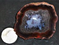
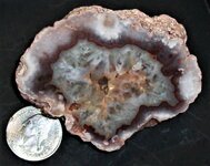
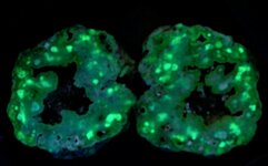
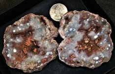
KT is certain some of you have heard of and perhaps seen Condor agates from the Andes Mountains, Mendoza Province, Argentina. There is also a different location for Puma agates, pseudomorphs after colonial coral, from Malargue City, Mendoza Province, Argentina. But have you ever put a LW or SW UV lamp light on them? Evidently there was a fair amount of Uranium in the water that formed them, for they are quite interesting in UV light! Typically more of a dull green in the banded segments and a brighter green in the crystalline (quartz) segments. Here is a matched pair of polished Puma agates, a single polished half of another, and a polished half of a Condor agate. KT picked these out for their different patterns of fluorescence.
The first pair of images is of the matched pair of Puma agates, US quarter for scale. The first picture is in natural light and the second is in SW 254nm, my 3 lamp setup. The third picture is of the single polished Puma half in natural light, with the same specimen in the fourth picture in SW 254nm light. The fifth picture is of a polished Condor agate, in natural light, US quarter for scale. The 6th picture is the same specimen in SW 254nm.
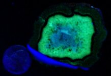
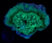
Then We have a micro specimen of Tyuyamunite on Selenite from the Qinglong Mine, Qinglong County, Guizhou Province, China. The Tyuyamunite crystals are scattered about on one surface of the specimen, having formed on a thin clearish crust of selenite in the first image. I was expecting to see the few scattered crystals in LW UV, but was shocked at what appeared under my USB led microscope at 25X! Apparently the selenite is also Uranium saturated for it fluoresces a solid green in LW 365nm light. Most unexpected, but since both these minerals are late forming in the fluid system, it stands to reason both may contain traces of U. His Majesty is quite happy about this specimen!
The first pair of images is of the matched pair of Puma agates, US quarter for scale. The first picture is in natural light and the second is in SW 254nm, my 3 lamp setup. The third picture is of the single polished Puma half in natural light, with the same specimen in the fourth picture in SW 254nm light. The fifth picture is of a polished Condor agate, in natural light, US quarter for scale. The 6th picture is the same specimen in SW 254nm.
Then We have a micro specimen of Tyuyamunite on Selenite from the Qinglong Mine, Qinglong County, Guizhou Province, China. The Tyuyamunite crystals are scattered about on one surface of the specimen, having formed on a thin clearish crust of selenite in the first image. I was expecting to see the few scattered crystals in LW UV, but was shocked at what appeared under my USB led microscope at 25X! Apparently the selenite is also Uranium saturated for it fluoresces a solid green in LW 365nm light. Most unexpected, but since both these minerals are late forming in the fluid system, it stands to reason both may contain traces of U. His Majesty is quite happy about this specimen!
Attachments
-
 Tyuyamunite with selenite, Qinglong Mine, Qinglong Co, Guizhou Province, China, 25X, LW 365nm.jpg278.5 KB · Views: 52
 -
 Tyuyamunite with selenite, Qinglong Mine, Qinglong Co, Guizhou Province, China, 25X, natural l...JPG191.7 KB · Views: 47
 -
 Condor agate, Andes Mountains, Mendoza Province, Argentina, US quarter for scale, SW 254nm.JPG75 KB · Views: 48
 -
 Condor agate, Andes Mountains, Mendoza Province, Argentina, US quarter for scale, natural light.JPG151.1 KB · Views: 50
 -
 Puma agate, ps. after coral, Malargue City, Mendoza Province, Argentina, US quarter for scale,...JPG81.7 KB · Views: 46
 -
 Puma agate, ps. after coral, Malargue City, Mendoza Province, Argentina, US quarter for scale,...JPG166.7 KB · Views: 49
 -
 Puma agate, matched pair, ps. after coral, Malargue City, Mendoza Province, Argentina, US quar...JPG63.4 KB · Views: 48
 -
 Puma agate, matched pair, ps. after coral, Malargue City, Mendoza Province, Argentina, US quar...JPG115.7 KB · Views: 49
While perusing eBay, KT saw a specimen that not only was within His Royal budget, but also was the best fluorescing specimen for its species that His Majesty had seen! JT bought it and it finally arrived today in the Royal Mailbox!
The specimen is Wollastonite and Hardystonite from the Desert View Mine, Holcomb Valley, San Bernardino Mountains, San Bernardino County, California. This site is known for several fluorescent minerals, including aragonite, calcite, hardystonite, willemite, and wollastonite, somewhat similar mineralogically to the Franklin, New Jersey, mineralization, although not as complex mineralogically.
The first picture is in natural light, with a US quarter for scale. The second picture is in SW 254nm, using the Royal 3 light set up. His Majesty is quite pleased with this specimen for its nice strong yellow response, and if you look closely there are a few patches of dark blue fluorescing Hardystonite, almost being wiped out by the strength of the Wollastonite’s response!
Enjoy!
Attachments
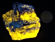
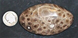
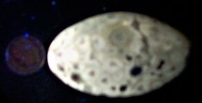
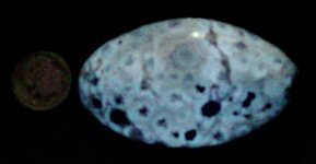
This fluorescent fossil arrived in the Royal Mailbox of the Castle today!
Here are 3 photographs of a polished example of colonial rugose coral that arrived today. Locally known as Petoskey Stone, it is actually called Hexagonaria percarinata, by paleontologists. It is the official state stone of Michigan. This example, which is tumble polished, came from a beach on Lake Michigan, in northern Michigan. Petoskey stones are composed predominately of calcite, which originated as an aragonite skeleton for the coral polyp, which apparently like colonial living, and with time inverted to the more stable carbonate...calcite. Typically each polyp’s home has a hexagonal structure, tightly packed with its neighbors in similar living conditions.
The first picture is in natural light, with a US dime for scale. The second picture is in LW 365nm and the fluorescence is strong enough to wipe out the skeletal details on the exposure meter of the Royal digital camera. The third picture is in SW 254nm from UV lamps, one at 3 o’clock and the other at 6 o’clock. The cell structure is better displayed in the 3rd picture.
Enjoy!
Attachments
-
 Coral, Petosky Stone, Lake Michigan beach, North Michigan, US dime for scale, natural light.JPG134.7 KB · Views: 49
 -
 Coral, Petosky Stone, Lake Michigan beach, North Michigan, US dime for scale, LW 365nm.JPG73.1 KB · Views: 52
 -
 Coral, Petosky Stone, Lake Michigan beach, North Michigan, US dime for scale, SW 254nm.JPG68.7 KB · Views: 46
The first specimen is from Mount Vesuvius, Italy. For the full location, read the picture label. It is a single crystal of Hauyne, a pale blue color. Since this mineral is within the Sodalite Group, it fluoresces a typical bright orange in LW 365nm. When this specimen arrived, it was mounted in a European T/N box, and when KT tried to remove it to mount in a more photographable orientation, the matrix crumbled in half! He checked both halves with the Royal LW UV lamp and fortunately, the crystal was still intact on the half that KT pulled out. The old tack had hardened so KT simply pulled it out of the box, tossed it and the poor half away. It was remounted properly for photography, and after several tries with the Royal Chinese toy microscope, IHis Majesty decided it was not being cooperative today, so the specimen went downstairs to the Wilde-Heerberg binocular scope and under 15X KT saw the best view. Both pictures taken using eye piece focus with the Royal digital camera. Picture 1 is in natural light and Picture 2 is in LW 365nm. The response is very bright, with orange light colorizing the entire inside of the base of the box.
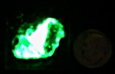
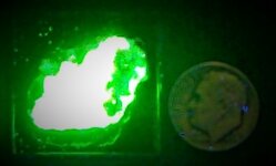
The second specimen is another brightly fluorescent mineral, a crust of Autunite, from Infesta Mine, Viariz, Baiao, Porto District, Portugal. This T/N's pictures were taken with the Royal digital camera at some 18 inches from the specimen and then the photo cropped to include the specimen and a US dime for scale. The first picture is in natural light, the second in SW 254nm (two lamps at 3 and 6 o'clock), and the third photo was taken with the Royal LW 365nm UVBeast at 3 feet and off center from the main beam. The green fluorescence in both LW and SW is so strong that it washed out the exposure meter on the digital camera and much of the most intense green fluorescence displays as white in the images.
Anyway, more fun and games! Enjoy the pictures!
Attachments
-
 Hauyne, Somma-Mount Vesuvius, Naples Prov., Campania, Italy, 15X, natural light.JPG191.4 KB · Views: 49
 -
 Hauyne, Somma-Mount Vesuvius, Naples Prov., Campania, Italy, 15X, LW 365nm.JPG75.1 KB · Views: 51
 -
 Autunite, Infesta Mine, Viariz, Baiao, Porto District, Portugal, US dime for scale, natural li...JPG107.7 KB · Views: 50
 -
 Autunite, Infesta Mine, Viariz, Baiao, Porto District, Portugal, US dime for scale, SW 254nm.JPG47.7 KB · Views: 49
 -
 Autunite, Infesta Mine, Viariz, Baiao, Porto District, Portugal, US dime for scale, LW 365nm.JPG72.4 KB · Views: 50
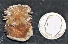
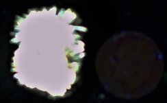
Here are 3 photos of a specimen of Schrockingerite. The specimen is from the Isolagrande Mine, Murialdo, Savona, Liguria, Italy. It is really a micromount with many crystals, but to view the overall fluorescence, it is better to view the entire specimen. Here are the photographs, all with a US dime for scale, so you could call the piece a small miniature, but the crystals are micros, tho some clusters are visible in the natural light photo.
Picture 1 is in natural light, picture 2 is in SW 254nm (KT's Royal three light setup), and picture 4 was taken in LW 365nm with the Royal UVBeast at 3 feet and out of the center of the beam! The green fluorescence is so intense that it wiped out the Royal Digital camera's exposure meter and shows as white in the strongest areas on the specimen.
Enjoy!
Attachments
-
 Schrockingerite crystals, Isolagrande Mine, Murialdo, Savona, Liguria, Italy, US dime for scal...JPG118.6 KB · Views: 48
 -
 Schrockingerite crystals, Isolagrande Mine, Murialdo, Savona, Liguria, Italy, US dime for scal...JPG71.3 KB · Views: 47
 -
 Schrockingerite crystals, Isolagrande Mine, Murialdo, Savona, Liguria, Italy, US dime for scal...JPG78.4 KB · Views: 46
A number of fluorescent specimens arrived today, in fact so many packages that the Royal mail delivery person left the door hanging wide open on the Royal mail box. Anyway, here are the first two specimens KT documented and photographed!
The first 3 pictures are of a Sodalite and feldspar specimen from an undisclosed quarry in the Tvedalin Region, Larvik, Vestfold og Telemark, Norway. There are some 15 or more quarries in that area, several of which contain sodalite according to Mindat.org, so that is as far as His Majesty could follow the mineralogy to get a location. The first picture is in natural light, and a US dime for scale. The second image is in LW 365nm, and the third picture is in SW 254nm. Typical strong orange coloration in LW.
The second specimen is of Aragonite, “strontian Aragonite”, also called Boccheggiano aragonite. From the Bocchegiano Mine, Montieri, Grosseto Province, Italy, likely the most famous location for fluorescent minerals in Italy. The first picture is in natural light, with a US dime for scale. Second picture is in LW 365nm, displaying the bright cherry red color typical of aragonite from this locality. The third picture is in SW 254nm, showing a subdued response of pale reddish orange.
Enjoy the pictures!
Attachments
-
 Sodalite & Feldspar, Undisclosed Qy., Tvedalen Region, Larvik, Vestfold og Telemark, Norway, U...JPG122.8 KB · Views: 47
 -
 Sodalite & Feldspar, Undisclosed Qy., Tvedalen Region, Larvik, Vestfold og Telemark, Norway, U...JPG76.4 KB · Views: 47
 -
 Sodalite & Feldspar, Undisclosed Qy., Tvedalen Region, Larvik, Vestfold og Telemark, Norway, U...JPG58.7 KB · Views: 45
 -
 Aragonite, Sr, Boccheggiano Mine, Montieri, Grosseto Prov., Italy, US dime for scale, natural ...JPG181 KB · Views: 53
 -
 Aragonite, Sr, Boccheggiano Mine, Montieri, Grosseto Prov., Italy, US dime for scale, LW 365nm.JPG93.6 KB · Views: 47
 -
 Aragonite, Sr, Boccheggiano Mine, Montieri, Grosseto Prov., Italy, US dime for scale, SW 254nm.JPG97.4 KB · Views: 47
This is the 2nd pair of specimens in yesterday’s shipment that filled the Royal Mailbox !
The first pair of photographs is of a Calcite from the Langban Mine, Langban Ore District, Filipstad, Varmland, Sweden. There are many documented locations at that mine, but the specimen label did not give any of those attributes. See this location listed in Mindat.org for further information. A US dime for scale is in each photograph. The first picture is in natural light and the second is in SW 254nm.
The next set of three photographs is of Sphalerite and Hydrozincite in Calcite, Hasselhojden Quarry, Bergslagen Mining District, Grythyttan, Hallefoks, Orebro Co., Sweden. Again a US dime for scale in each picture. The first picture is in natural light, the second is in LW 365nm with the sphalerite luminescing gold and hydrozincite showing up as blue-white spots scatter about. The third picture is in SW 254nm, the calcite displaying red fluorescence.
SW images in both cases were taken using the Royal 3 SW lamp set up in typical format.
Enjoy the pictures!
Attachments
-
 Calcite, Langban Mine, Langban Ore Distr., Filipstad, Varmland, Sweden, US dime for scale, nat...JPG162.5 KB · Views: 51
 -
 Calcite, Langban Mine, Langban Ore Distr., Filipstad, Varmland, Sweden, US dime for scale, SW ...JPG83.4 KB · Views: 49
 -
 Sphalerite & Hydrozincite in Calcite, Hasselhojden Qy., Bergslagen Min. Distr., Grythyttan, Ha...JPG170.5 KB · Views: 45
 -
 Sphalerite & Hydrozincite in Calcite, Hasselhojden Qy., Bergslagen Min. Distr., Grythyttan, Ha...JPG151.7 KB · Views: 52
 -
 Sphalerite & Hydrozincite in Calcite, Hasselhojden Qy., Bergslagen Min. Distr., Grythyttan, Ha...JPG79.6 KB · Views: 47
Well, the Royal Mailbox is being kept busy! 
One package arrived today from Spain and it contained two interesting specimens.
The first 3 pictures are of a double grouping of acicular Aragonite needles from La Tinaja Quarry, La Serna, Pantoja, Toledo, Castile-La Mancha, Spain. US dime for scale. The first picture is in natural light. The second is in LW 365nm. The LW response is such an intense white it wiped out the Royal digital camera’s exposure meter, hence the white blob in the 2nd photo. The third picture is in SW 254nm (3 lamp set up).
The second specimen is a single clear selenite crystal, a variety of gypsum. It is from Pomeno, Chelva, Valencia Community, Spain. It was not being described as a fluorescent mineral, but previous examples from various localities indicate these single gypsum crystals often display a fluorescent hourglass pattern. When they do, most of the time they formed in black organic clay beds. KT took a chance and ordered this one just to see! The first picture is in natural light and the second is in LW 365 (the Royal UVBeast torch held some 4 feet away and off the center of the beam). We see this same hourglass pattern of tan sand in the selenite crystals of the Great Salt Plain, near Jet, Oklahoma. KT thinks it perhaps relates to the fact that some crystal faces have a greater crystallographic strength during growth and can push grains aside, where as faces with weaker crystallographic strength cannot and therefore are highly included. Anyway, single gypsum crystals with this form are among my Royal favorites!
Enjoy the pictures!
Attachments
-
 Aragonite,La Tinaja Quarry, La Serna, Pantoja, Toledo, Castile-La Mancha, Spain, US dime for s...JPG108 KB · Views: 50
 -
 Aragonite,La Tinaja Quarry, La Serna, Pantoja, Toledo, Castile-La Mancha, Spain, US dime for s...JPG65.3 KB · Views: 47
 -
 Aragonite,La Tinaja Quarry, La Serna, Pantoja, Toledo, Castile-La Mancha, Spain, US dime for s...JPG53 KB · Views: 48
 -
 Gypsum, var. selenite, Domeno, Chelva, Valencia Community, Spain, US dime for scale, natural l...JPG121.9 KB · Views: 47
 -
 Gypsum, var. selenite, Domeno, Chelva, Valencia Community, Spain, US dime for scale, LW 365nm.JPG69.9 KB · Views: 47
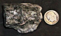
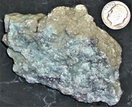
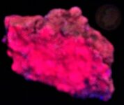
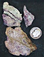
While rearranging the Royal Mineral Collection to make room for a 6th LW drawer in KT's Collector Cabinet, His Majesty came across these specimens that He had collected years ago as part of a suite of samples from His Master’s thesis area. They came from the type locality of Miserite. The Arkansas Novaculite, a high silica rock, was invaded by fluids from a Carbonatite, and what KT termed in His thesis as a reverse skarn was formed. Normally skarns are formed from the invasion of limestone by silica-rich fluids coming off a granitic magma and causing alteration to the limestone host rock. During His thesis work, KT actually found a zone of recrystallized Novaculite, that displayed a chemical reaction front frozen in it where wollastonite had been forming from fluids evolving from a nearby carbonatite. Miserite is a chemical sponge mineral, capable of accommodating many different elements during its formation.
Anyway, these specimens are from that alteration zone on the east side of the North Wilson pit, Potash Sulphur Springs, Garland County, Arkansas. The mineral was first analyzed by the Chem Lab of the State Geologist’s Office in the early 1890s and called Natroxontolite, but later reanalyzed by Waldmar Schaller and discovered that what was thought to be Sodium in the structure is actually Calcium. Schaller renamed it Miserite, in honor of H. D. Miser, early Arkansas educated geologist, who produced the first Geological Map of Arkansas in 1929. Later work by a crystallographer determined that this mineral is the first known species to fit the theoretical structure termed Zoltai 5. This effort was aided by the discovery of actual crystals of Miserite from Mont St. Hilaire in Canada.
The first picture is in natural light, the Miserite is scaly in habit, pink in color, and intimately intergrown with wollastonite. US dime for scale. The second picture shows the yellow response of the miserite to SW 254nm in a two lamp exposure, one at 3 o’clock and the other at 9 o’clock. At some time in the past, KT had collected and labeled these specimens as SW fluorescent, having checked them with His old 120V UV Products SW lamp. Interestingly the wollastonite is not responsive to either SW or LW UV. KT knows these specimens have been in his possession over 30 years!
Enjoy the pictures.
Attachments
After arriving home today from lunch with friends, KT found The Royal Mailbox jammed with packages….again! So His Majesty has 3 specimens for your examination today.
The first pair of pictures are of calcite and sulfur from the Cianciana Sulfur Mine, Agrigento Province, Sicily, Italy. This specimen is definitely a cabinet piece as KT had to use an old Peace silver dollar for scale! The first picture is in natural light and the second is in LW 365nm. The specimen to the eye is a bit brighter than the picture in LW.
The second specimen is another calcite from the Monte Arsiccio Mine, Stazzena, Luccia Province, Tuscany, Italy. It is a fist size cluster of cave type calcite. A US quarter for scale. The first picture is natural light and the second is LW 365nm. Interesting white with a hint of bluish tinge.
The third specimen is a sample of tremolite in marble from Ziffertal, Schwaz District, Tyrol, Austria. US dime for scale. The first picture is in natural light and the second in LW 365nm. Any white in the LW image is a blowout of the Royal digital camera’s meter, as it should all appear strongly fluorescent but pale blue in color.
Enjoy the pictures.
Attachments
-
 Calcite & Sulfur, Cianciana Mine, Agrigento Province, Sicily, Italy, US silver dollar for scal...JPG96 KB · Views: 41
 -
 Calcite & Sulfur, Cianciana Mine, Agrigento Province, Sicily, Italy, US silver dollar for scal...JPG88.3 KB · Views: 45
 -
 Calcite, Monte Arsiccio Mine, Stazzena, Lucca Province, Tuscany, Italy, US quarter for scale, ...JPG198.6 KB · Views: 44
 -
 Calcite, Monte Arsiccio Mine, Stazzena, Lucca Province, Tuscany, Italy, US quarter for scale, ...JPG139.8 KB · Views: 47
 -
 Tremolite, Zillertal, Schwaz District, Tyrol, Austria, US dime for scale, natural light.JPG136.5 KB · Views: 48
 -
 Tremolite, Zillertal, Schwaz District, Tyrol, Austria, US dime for scale, LW 365nm.JPG124.1 KB · Views: 43
GKL
Forum Supporter
Sorry for so long in between posting, I don't mean to go so long between posts, it just seems like I've been keeping busier than usual this year and I realize it's been longer than I intended since I posted.
Anyhow KT, you never seem to fail to amaze me with how many neat new specimens you manage to get from the previous time I check, I appreciate that you take the time to post the photos along with interesting info about them, and there seems to be so many minerals I never heard of before so it is definitely educational too !
I now wish they had a class just on fluorescent minerals when I went to high school
When KT went to high school lo so many years ago, they taught nothing about mineralogy....Heck, His Majesty had to go to college to take a mineralogy course and then KT does not even remember fluorescence being mentioned!Sorry for so long in between posting, I don't mean to go so long between posts, it just seems like I've been keeping busier than usual this year and I realize it's been longer than I intended since I posted.
Anyhow KT, you never seem to fail to amaze me with how many neat new specimens you manage to get from the previous time I check, I appreciate that you take the time to post the photos along with interesting info about them, and there seems to be so many minerals I never heard of before so it is definitely educational too !
I now wish they had a class just on fluorescent minerals when I went to high school
That is one reason that KT likes to give talks to the geology students....simply because frequently fluorescence is a property overlooked when trying to identify minerals. And they really need to be aware of its existence!
Speaking of existence, today His Majesty received one fluorescent specimen in the Royal Mailbox so this is a good place to show it! It is Meionite, a variety of Scapolite with non-fluorescent Diopside crystals. The piece is from Slyndyanka, Lake Baikal, Irkutsk Oblast, Russia. The specimen has been acid cleaned to remove calcite and display the diopside crystals, but that process also exposed the Scapolite that composes part of the matrix.
The first picture is in natural light with a US dime for scale. The second picture was taken in LW 365nm light.
Enjoy the pictures! And many various specimens are on their way to the Castle, so KT patiently awaits their arrival.

Attachments
A visitor to the Royal Castle bearing gifts for KingTotsalot last night and a Royal Journey today!
KT's visitor was a fellow rockhound who was on his way back to the East Coast of the US when he decided to stop in and present His Majesty with several fluorescent specimens that he had collected on his trip out west! One of these is featured in the first pair of pictures!
The small community of Mt. Ida in Montgomery County, some 2 hour drive west of KT's Castle, was hosting a rock and mineral show and KT decided to attend. Even though it was a bright sunny day, His Majesty managed to find two nice fluorescent specimens at bargain prices so He brought them back for the Royal Collection.
The first pair of pictures is of Chalcedony, a microfibrous variety of quartz, from the North Slope, Saddle Mountain, west of Phoenix, in Maricopa County, Arizona. A US dime is shown for scale in the photos. The first image is in natural light, and the second is in LW 365nm. The green coloration in UV is due to traces of Uranium incorporated in the mineral as it formed.
The second pair of pictures is of Sodalite with Lapis. There have been active Lapis mines in Kokcha Valley, Badakhshan, Afghanistan for over 6,000 years! Again a US dime for scale. The first picture is in natural light and the second shows the orange fluorescing sodalite within the vein of Lapis in LW 365nm.
The final pair of pictures are of a specimen of greenish fluorite from Hunan Province, China. US quarter for scale. The first picture is natural light and the second is in LW 365nm. The entire face should appear blue, but the Royal Camera's photo meter was overcome by the intensity in the upper portion of the picture, causing it to look white from the washout.
So KT enjoys being out with his peoples and was given all due respect by them! Even to the point that his meal and fuel for his horses were paid for on this trip! There are more pictures from KT's guest specimens to come as His Majesty has time to sort through them!
Enjoy!
Attachments
-
 Chalcedony, North Slope, Saddle Mountain, West of Phoenix, Maricopa Co., AZ, US dime for scale...JPG142.2 KB · Views: 36
 -
 Chalcedony, North Slope, Saddle Mountain, West of Phoenix, Maricopa Co., AZ, US dime for scale...JPG96.7 KB · Views: 45
 -
 Sodalite with Lapis, Kokcha Valley, Badakhshan, Afghanistan, US Dime for scale, natural light.JPG163.7 KB · Views: 45
 -
 Sodalite with Lapis, Kokcha Valley, Badakhshan, Afghanistan, US Dime for scale, LW 365nm.JPG105.5 KB · Views: 33
 -
 Fluorite, Hunan Province, China, US quarter for scale, natural light.JPG208.4 KB · Views: 49
 -
 Fluorite, Hunan Province, China, US quarter for scale, LW 365nm.JPG104.8 KB · Views: 43