One more specimen from the large box.
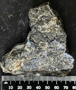
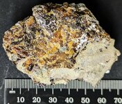
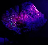
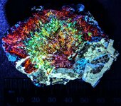
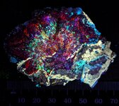
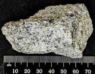
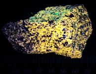
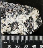
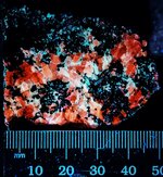
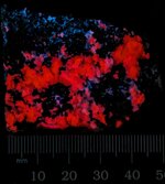
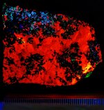
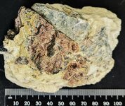
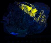
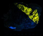
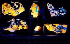
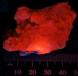
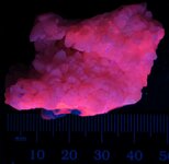
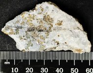
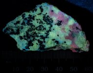
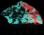
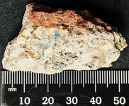
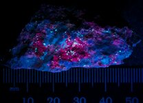
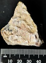
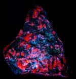
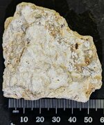
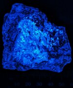
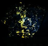
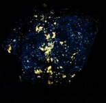
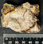
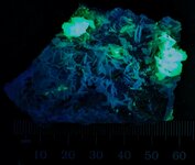
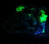
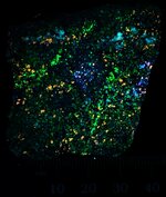
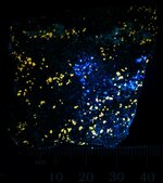
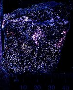
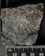
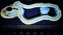
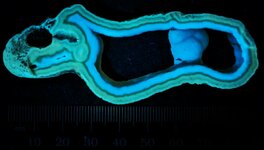
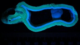
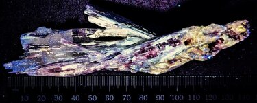
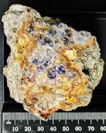
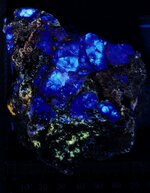
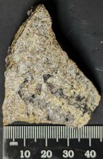
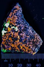
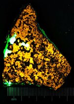
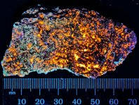
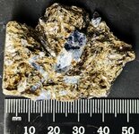
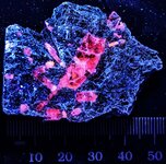
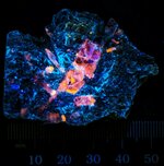
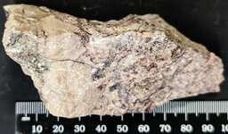
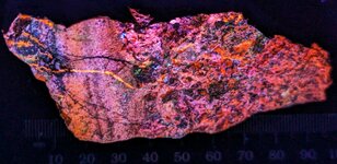
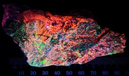
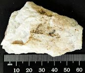
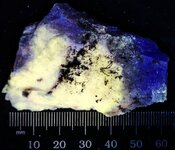
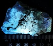
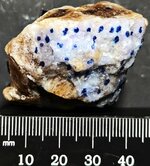
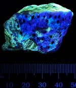
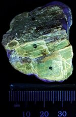
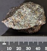
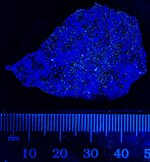
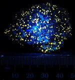
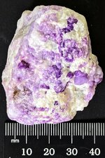
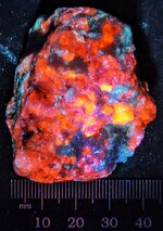
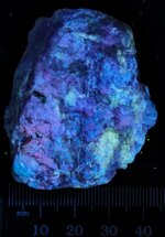
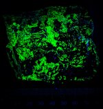
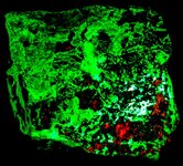
This cabinet sized specimen consists of fluorescent Willemite, Calcite, and Fluorapatite with Andradite, Hendricksite, Rhodonite, and Franklinite from the Franklin Mine, Franklin, Sussex Co., NJ.
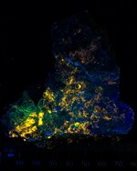
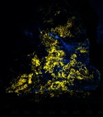
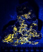
The first picture is in natural light. The second picture is in LW 365nm with Willemite predominating. The third picture is in SW 254nm with Willemite responding green, Calcite as scattered red spots, and Fluorapatite responding as scattered area of pinkish white.
Enjoy the pictures!
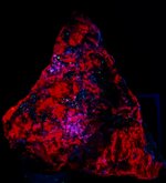
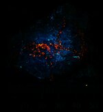
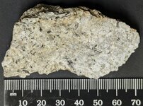
This cabinet sized specimen consists of fluorescent Willemite, Calcite, and Fluorapatite with Andradite, Hendricksite, Rhodonite, and Franklinite from the Franklin Mine, Franklin, Sussex Co., NJ.
The first picture is in natural light. The second picture is in LW 365nm with Willemite predominating. The third picture is in SW 254nm with Willemite responding green, Calcite as scattered red spots, and Fluorapatite responding as scattered area of pinkish white.
Enjoy the pictures!
Attachments
-
 Willemite, calcite, fluorapatite, & others, Franklin Mine, Franklin, Sussex Co., NJ, natural l...jpg331.9 KB · Views: 16
 -
 Willemite, calcite, fluorapatite, & others, Franklin Mine, Franklin, Sussex Co., NJ, LW 365nm.jpg145.9 KB · Views: 11
 -
 Willemite, calcite, fluorapatite, & others, Franklin Mine, Franklin, Sussex Co., NJ, SW 254nm.jpg229 KB · Views: 13